Imaging Systems
X-ray image intensifier
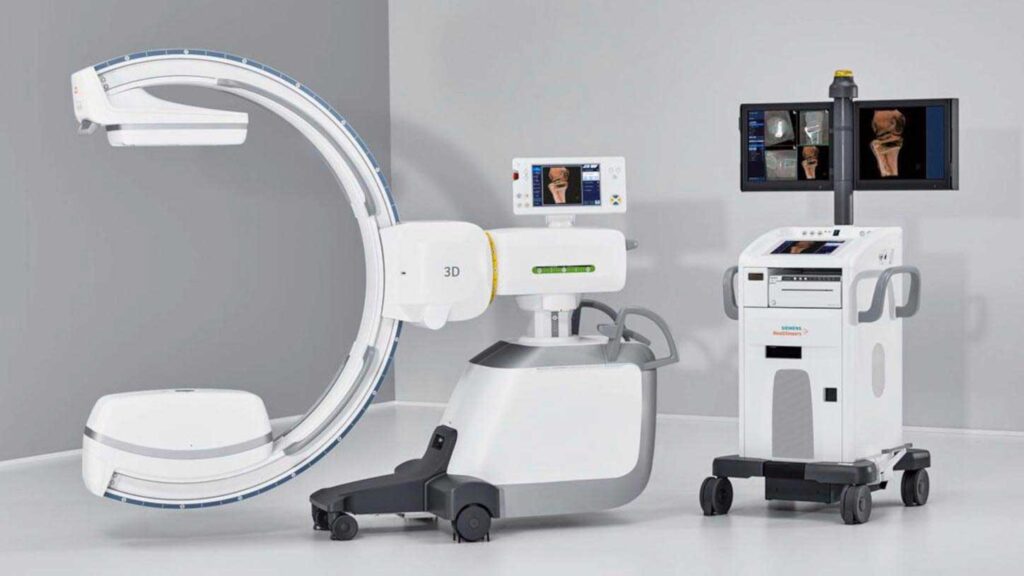
An X-ray image intensifier (XRII) is an image intensifier that converts X-rays into visible light at higher intensity than the more traditional fluorescent screens can. Such intensifiers are used in X-ray imaging systems (such as fluoroscopes) to allow low-intensity X-rays to be converted to a conveniently bright visible light output. The device contains a low absorbency/scatter input window, typically aluminum, input fluorescent screen, photocathode, electron optics, output fluorescent screen and output window. These parts are all mounted in a high vacuum environment within glass or more recently, metal/ceramic. By its intensifying effect, It allows the viewer to more easily see the structure of the object being imaged than fluorescent screens alone, whose images are dim. The XRII requires lower absorbed doses due to more efficient conversion of X-ray quanta to visible light.

Full-field Digital Mammography Unit
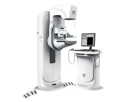
ASR-4000 digital mammography system can fully satisfy the requirements of current breasts imaging. It can be equipped with minimally invasive biopsy device with threedimensional positioning technology, which provides imageguided stereotactic to improve the accuracy and detectable rate, as a result, it improves the diagnosis and gives better support in choosing reasonable treatment plan. It can realize cone beam computed tomography (CBCT) which can solve the problem about calcify points covered by gland under 2D photography, hence, it improves the clinical diagnosis. ASR-4000 digital mammography system is a perfect combination between the advanced detector technology and dedicated x-ray tube for breast examination. It adopts the third generation full-size amorphous selenium flat-panel detector, converting the x-ray to digital signal directly, therefore, it ensures high x-ray conversion efficiency and higher image resolution with over 51p/mm, it helps to find out tiny calcification at the early stage of breast cancer. Compared to last two generations of amorphous selenium detecting technology, the third generation technology is better in integration, acquisition rate and transmission rate, besides, it has better adaptability to the environment, for instance, the temperature and humidity. Compared to traditional molybdenum target mammography, the third generation amorphous selenium digital breast imaging adopts brand-new optical conversion technology with low noise, high resolution and high x-ray conversion efficiency, it allows low dosage and ensures high-quality image.

